
In this week's edition
- ✍️ Letter from P'Fella
Designer Nipples... - 🤓 The Sunday Quiz
How well do you know the facial nerve? - 🖼️ Image of the Week
Signs of facial nerve palsy. - 🚑 Technique Tip
Sural nerve graft location for nerve recon. - 📖 What Does the Evidence Say
Facial nerve branch localization. - 🔥 Articles of the Week
Facial nerve danger zones, parotidectomy, & assessing the facial nerve: with 1 sentence summaries. - 💕 Feedback
Suggest ideas & give feedback!
A Letter from P'Fella
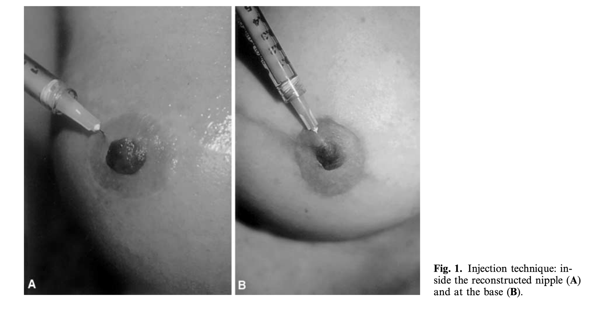
Designer Nipples....
Nothing is currently off limits—even our most unexpected features. We have evolved from cheek and lip fillers to a trend that now offers a quick shot of hyaluronic acid to enhance nipple appearance. Priced ~$5K and promising results from six months to two years, this development is a perky twist...
Before you raise an eyebrow, consider this: a 2017 Independent article already spotlighted “designer nipples” as a controversial trend, highlighting how the quest for perfection isn’t just about major features. Scientific literature has documented the evolution of nipple reconstruction and augmentation techniques. So, are we truly witnessing innovation, or is this just an old idea repackaged for today’s market?
The Big Question
Here’s where it gets controversial: Is this a genuine step toward personal empowerment, or are we just enabling an industry that thrives on our insecurities? This isn’t about passing judgment on anyone’s body— the literature shows all genders have reported outcomes - it’s about the fine line between self-expression and a market-driven obsession with perfection.
Downstream Effects
I'm particularly interested in the second-order effects. Redesigning every detail creates ripple effects throughout our practice. For example, altering nipple anatomy may complicate breast screening and imaging, while changes in tissue contours and projections can obscure subtle signs on mammograms or ultrasounds, potentially leading to diagnostic challenges later. Even minor cosmetic procedures can have significant consequences for patient care.
With love,
P'Fella ❤️
The Sunday Quiz
How Well Do You Know the Facial Nerve?
Welcome to the next round of The Weekly Quiz.
Each edition of thePlasticsPaper includes a quiz question designed to challenge and engage our readers. Keep your wits about you and join in every week — the winner at the end of six rounds will earn you a one-year subscription to thePlasticsPro.
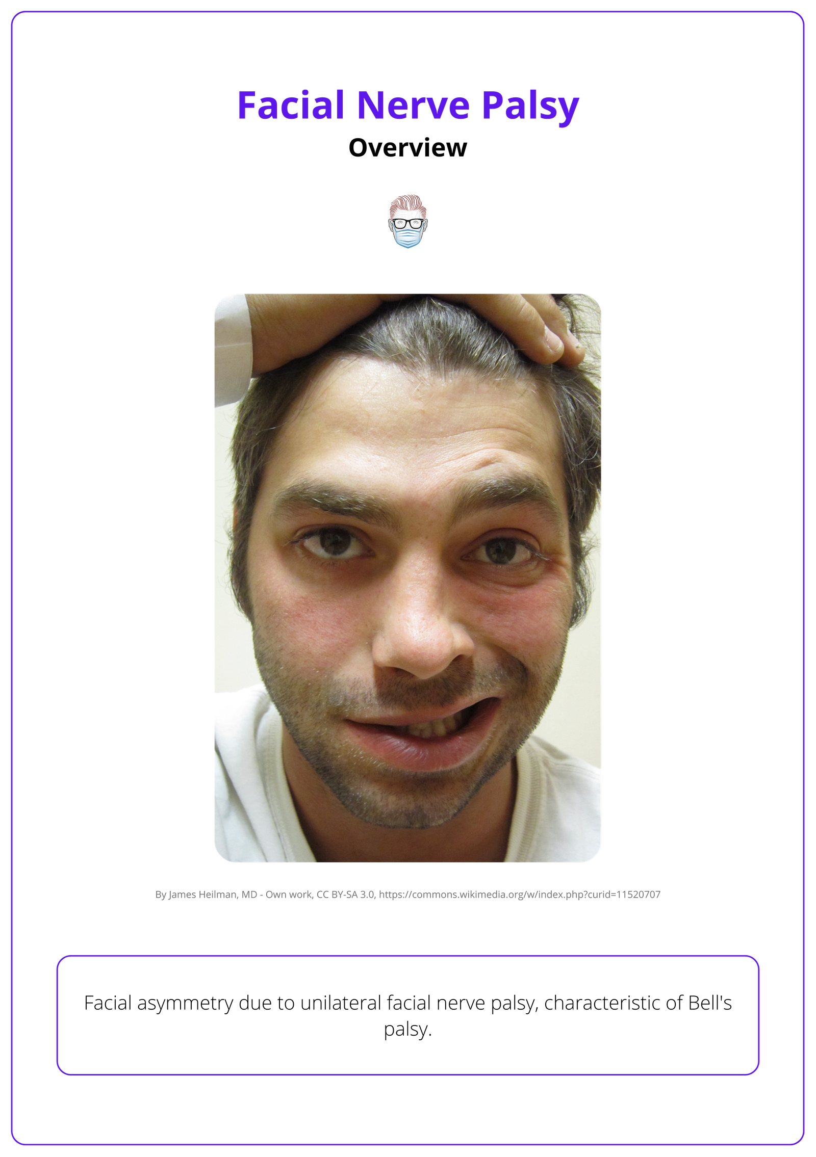
Image of the Week
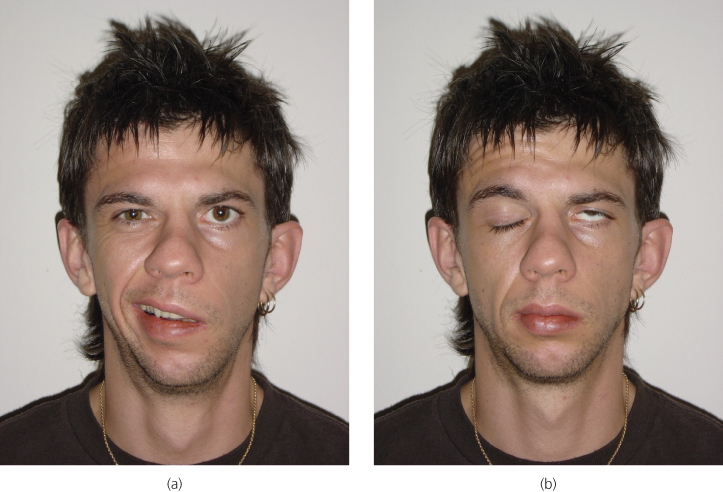
Signs of Facial Nerve Palsy
In this section, we feature an anatomical illustration. This week’s image highlights left-sided lower motor neuron facial nerve palsy.
(A) Generalized muscle weakness affecting the forehead, cheek, and mouth.
(B) Positive Bell’s phenomenon — upward eye movement when attempting to close the left eye.
Technique Tip
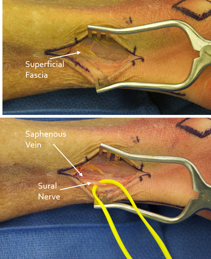
Sural Nerve Graft Location
This week, we’re focusing on optimal sural nerve graft location for nerve reconstruction.
Landmarks: Found beneath superficial fascia, adjacent to the saphenous vein.
Technique: Incise over the posterolateral ankle, dissect carefully to expose the nerve.
Tip: Preserve the saphenous vein to minimize trauma.
Efficient dissection ensures a viable graft for reconstruction.
What Does the Evidence Say?
Facial Nerve Branch Localization
Several landmarks have been proposed to locate the nerve, including the frontal branch of the superficial temporal artery (Lei et al., 2005) and a point 3.21 cm from the tragus along the zygomatic arch (Sanderson et al., 2020). The nerve is generally found within 2.5-3 cm of the lateral orbital rim and 2.85 cm superior and 2.54 cm lateral to the lateral canthus (Hashmi et al., 2021). Various surgical techniques have been developed to protect the nerve during procedures, including interfascial dissection and combined frontotemporal scalp/superficial temporal fascia dissection (Pekár et al., 2004).
Articles of the Week
3 Interesting Articles with 1 Sentence Summaries
Understanding the three-dimensional anatomy and danger zones of the facial nerve is crucial for preventing iatrogenic injury during facial rejuvenation surgery.
Malignant parotid tumors often necessitate facial nerve sacrifice due to locoregional invasion, increasing the need for microsurgical reconstruction, nerve grafting, or reinnervation to optimize outcomes.
Facial nerve palsy management depends on duration, location, and mechanism of injury. Early cases may allow direct repair, grafting, or nerve transfer, while prolonged palsy requires free functional muscle transfer for reanimation.
